


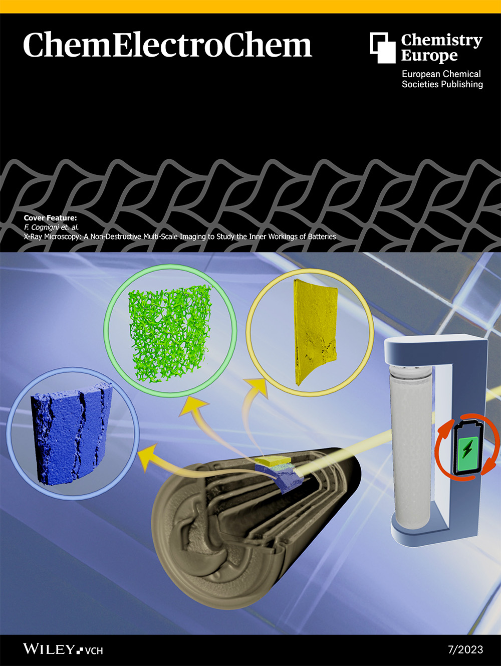
The leading scientific journal "ChemElectroChem" dedicates the cover of its n. 7 issue to a research entitled "X-Ray Microscopy: A Non-Destructive Multi-Scale Imaging to Study the Inner Workings of Batteries," carried out as part of the iENTRANCE project, at the new CNIS laboratories in Sapienza. Authors: Dr. Flavio Cognigni, Professor Mauro Pasquali, Dr. Francesca Anna Scaramuzzo, and Professor Marco Rossi of ICI-SBAI.
"X-ray microscopy (XRM) is a non-destructive characterization technique that provides quantitative information regarding the morphology/composition of the specimen and allows to perform multiscale and multimodal 2D/3D experiments exploiting the radiation-matter interactions. XRM is particularly suitable to afford in situ images of inner parts of a battery and for the early diagnosis of its degradation in a non-invasive way. Since traditional characterization techniques (SEM, AFM, XRD) often require the removal of a component from the encapsulated device that may lead to non-desired contamination of the sample, the non-destructive multi-scale potential of XRM represents an important improvement to batteries investigation. In this work, we present the advanced technical features that characterize a sub-micron X-ray microscopy system, its use for the investigation of hidden and internal structures of different types of batteries and to understand their behavior and evolution after many charge/discharge cycles."
Full article in:
https://chemistry-europe.onlinelibrary.wiley.com/doi/full/10.1002/celc.202201081?fbclid=IwAR1aB5FQx5rvmYV_2Ol7V9kazp1mGnSVjmUCWOiv29CXVQGuTuXu7isVdYE